Diagram Of The Eye Front View

Anatomy Human Eye Front View External Stock Illustration
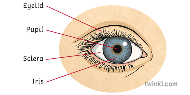
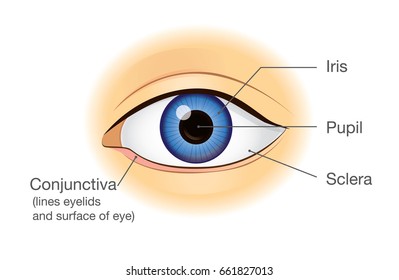
Diagram Of The Eye Front View Illustration Twinkl

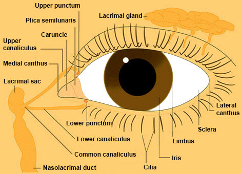
Eye Structures Front And Side Views Healthlink Bc
Human eye anatomy structure and function.
Diagram of the eye front view. Schematic diagram of the structure of the human eye. As a bonus site members have access to a banner ad free version of the site with print friendly pages. The eye one of the most complex organisms in the human body. The front part what you see in the mirror includes.
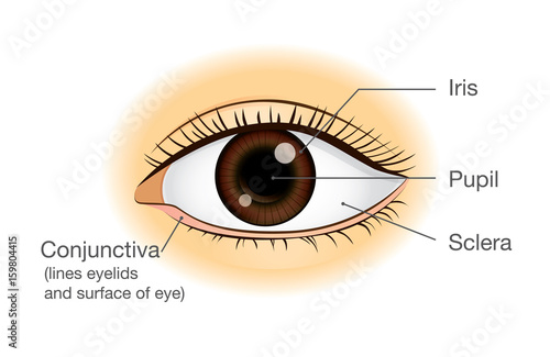
Is the most valuable and sensitive sense organ and it is a natural optical instrument. The inner sensorineural layer is known as the retina. It can open or close to widen or narrow the central aperture known as the pupil. A closer look at the parts of the eye by liz segre when surveyed about the five senses sight hearing taste smell and touch people consistently report that their eyesight is the mode of perception they value and fear losing most.
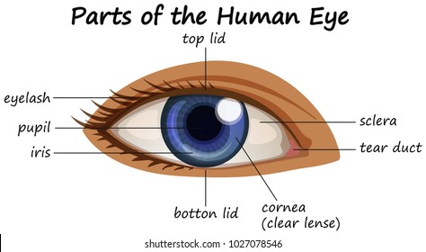
Cornea iris pupil eye lens retina. Draw a diagram of the human eye as seen in a vertical section and label the parts which suits the following descriptions relating to the. The cornea is a strong clear bulge located at the front of the eye where it replaces the. Diagram of the front view of human eye.
This simple introduction the subjects of the eye and visual optics includes a simple diagram of the eye together with definitions of the parts of the eye labelled in the illustration. Just behind the iris and pupil lies the lens which helps focus light on. The eye diagram is used primarily to look at digital signals for the purpose of recognizing the effects of distortion and finding its source. Parts of human eye with name stock vector illustration image.
The important parts of the eye. In order for the eye to work at its best all parts must work well collectively. In this manner it can control the amount of light entering the eye. The eye diagram takes its name from the fact that it has the appearance of a human eye.
Diagram human eye blind spot with eye anatomy of the human eye in front view external schematic diagram detailed anatomy of the human eye. It is made up of many different parts working in unison together. Asked jan 9 in class x science by navnit40. Related questions 0 votes.
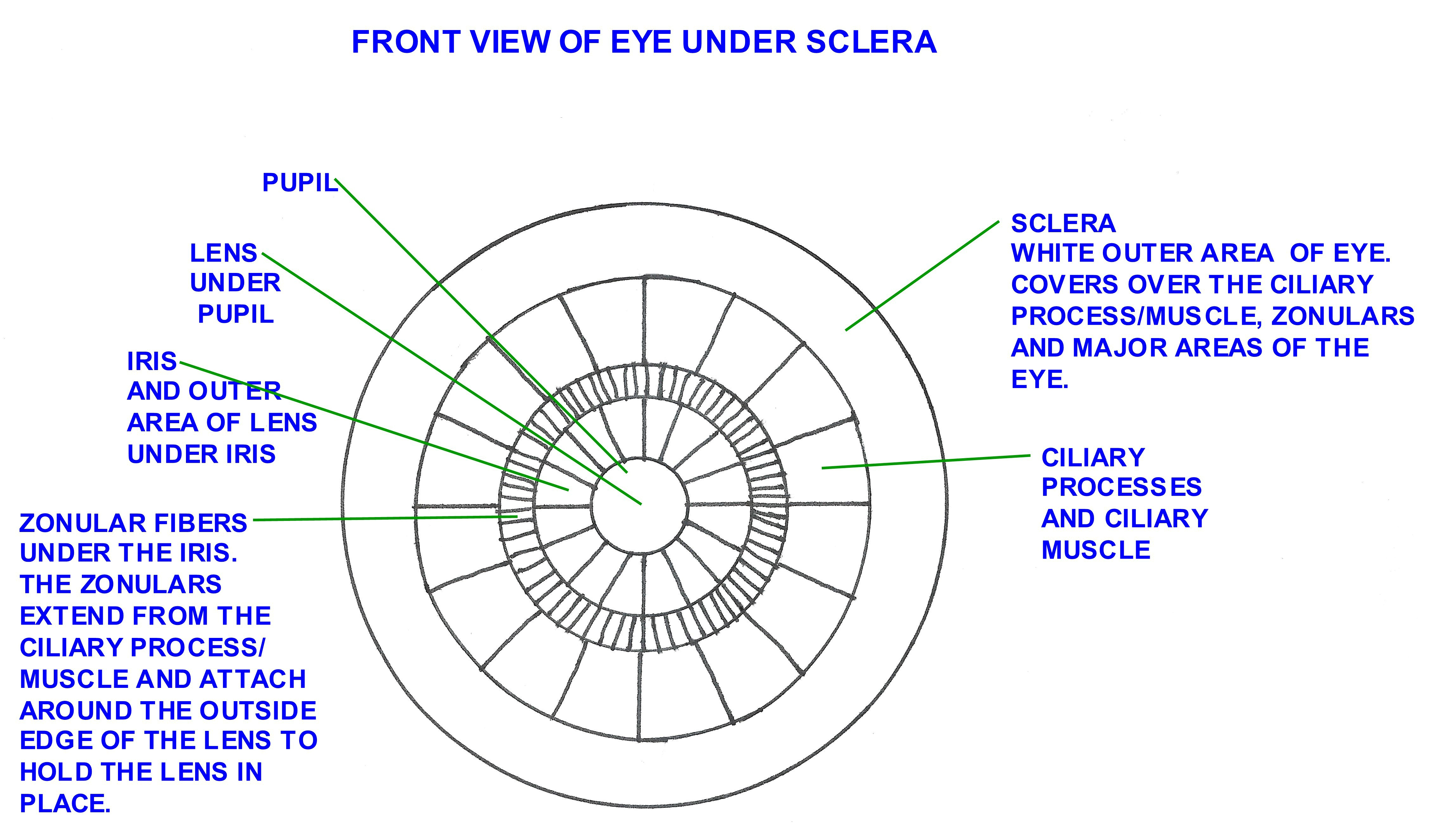
Diagram of the choroid iris and ciliary body. It is created simply by superimposing successive waveforms to form a composite image.

Front View Of A Human Eye Download Scientific Diagram

Front View Of The Human Eye Download Scientific Diagram

Draw A Labeled Diagram Of The Front View Of Human Eye

Anatomy And Physiology Chapter 10 Eye Front View Diagram
Similar Images Stock Photos Vectors Of Human Eye Anatomy
Human Eye Anatomy In Front View Illustration About Physical

Discover All About The Eye Its Anatomy Its Functioning

Front View Of The Human Eye Download Scientific Diagram
Eye Health Anatomy Of The Eye Visionaware

Eye Anatomy Page 13

Eye Structures Front And Side Views Healthlink Bc
Gallery View Anatomy The Eyes Have It

Front View Of The Eyelids Pupil And Other Visible Parts Of
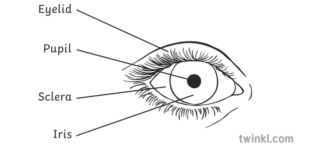
Diagram Of The Eye Front View Black And White Illustration

Normal Anterior Eye Front View And Diagram Photo Left
Human Eye Eye Changes Length Like A Camera To Focus Close

Anatomy Of The Eye Doctor Stock
Similar Images Stock Photos Vectors Of Bacterial
