Diagram Of An Onion Epidermal Cell

Cbse Class 9 Science Practical Skills Slide Of Onion Peel

Microscope Cell Lab Cheek Onion Zebrina Schoolworkhelper
Mic Uk

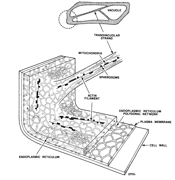
It is surrounded by cytoplasm.
Diagram of an onion epidermal cell. An onion is made up of layers that are separated by a thin membrane. Although at the surface an onion has dried protective leaf inside of it things change. Visit the post for more. The onion cell diagram shows that in the middle of the vegetable there is fairly much water.
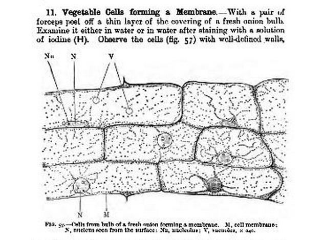
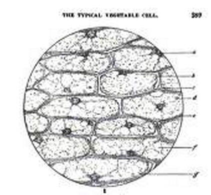
As in all plant cells the cell of an onion peel consists of a cell wall cell membrane cytoplasm nucleus and a large vacuole. An epidermal onion cell diagram includes components such as citoplasm a round nucleus and a cell wall. Easily obtained inexpensive they offer samples with no difficult technique required. The cell diagram displays a hexagonal shape of the epidermal onion cells and the way they are clustered.
These large cells from the epidermis of a red onion are naturally pigmented. The nucleus is present at the periphery of the cytoplasm. While both onion and human cells have a cell membrane only the onion has a cell wall. The microscope is the device which helps scientists observe how the onion epidermal layer looks like.
In this lab students will be able to observe the following organelles. Unlike animal cells such as cheek cells the cell wall of an onion and other plants are made up of cellulose which protects the cell and maintains its shape. 2 marks ans 44. The epidermal cells of onions provide a protective layer against viruses and fungi that may harm the sensitive tissues.
For this experiment the thin membrane will be used to observe the onion cells. The thin layer of skin found on the inside of an onion scale one layer of onion lifts off without effort and can be wet mounted on a slide with no need for extreme skill. Draw a labelled diagram of an onion epidermal cell seen under the microscope. Both plant and animal cells including human epithelial and onion epidermal cells have a structure called a cell membrane or plasma membrane.
Onion cell microscope drawing snc2dmrscott now let s view our slide the diagram below shows an onion cell taken from bulb of and a red blood is special animal that does not the image in left is a schematic diagram of onion epidermal cells plated with. Because of their simple structure and transparency they are often used to introduce students to plant anatomy 1 or to demonstrate plasmolysis. Iodine and methylene blue are required for this experiment and are common in any schools science la. 4 marks e the onion epidermal cells are not green in colour because they lack an organelle.
Nucleus cell wall cytoplasm and the nucleolus within the cells of an onion and a cheek. An onion is a multicellular plant organism. Onion and cheek cell lab experiment.
Microscope Cell Lab Cheek Onion Zebrina Schoolworkhelper
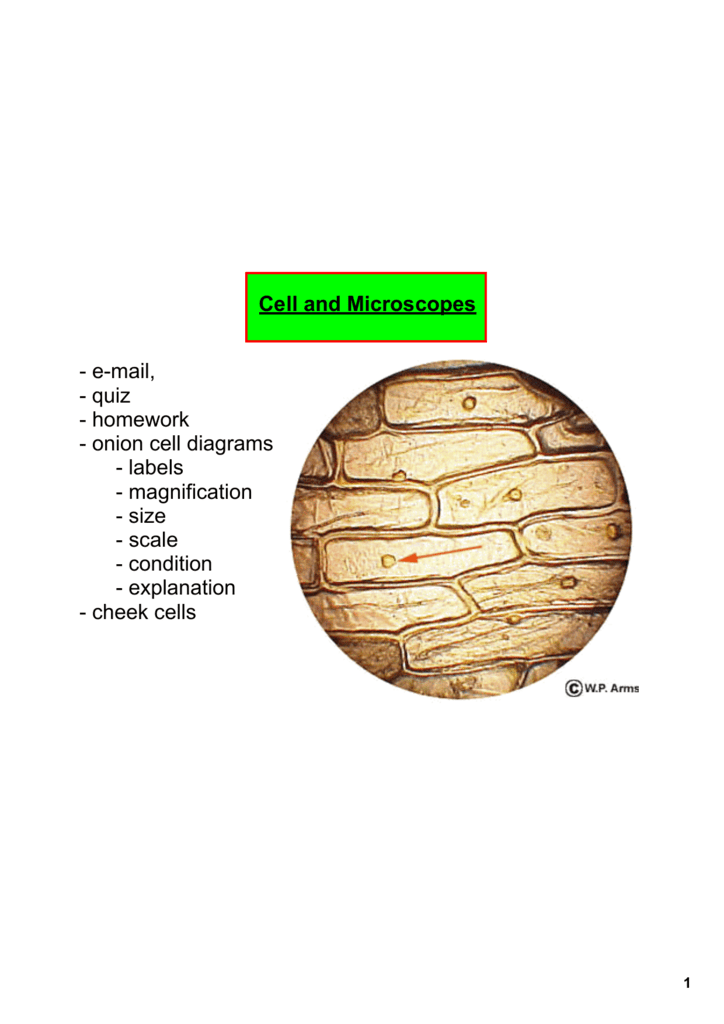
Cell And Microscopes Email Quiz Homework Onion Cell Diagrams

Cbse Class 9 Science Practical Skills Slide Of Onion Peel

Labeled Onion Cell Epidermis Cell Wall Cell Membrane Onion

Microscopy How A Microscope Works Magnification Calculations
Mic Uk
Practical Booklet Biology4isc

Ncert Class 9 Science Lab Manual Slide Of Onion Peel And
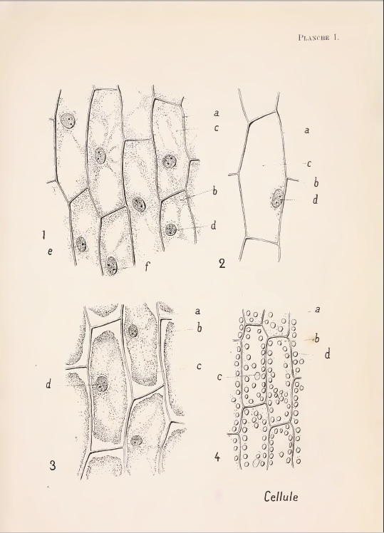
Diagram Of Onion Epidermal Cell Wiring Diagrams Folder

14 Best Scientific Drawing Images Scientific Drawing
Mic Uk The Inner Epidermis Of The Onion Bulb Cataphylls

Cbse Class 9 Science Practical Skills Slide Of Onion Peel

Epidermal Onion Cells Under A Microscope Plant Cells Appear

Labeled Onion Cell Diagram Wiring Diagrams Simple

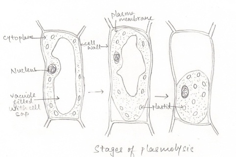
Observing Osmosis Plasmolysis And Turgor In Plant Cells

Schematic Of Experimental Set Up For Onion Epidermis Cell

Microscope Cell Lab Cheek Onion Zebrina Schoolworkhelper

Scientists Create Artificial Muscles From Gold Onion
